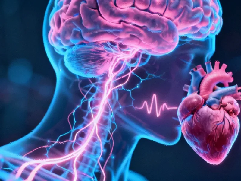
The Brain-Heart Communication Pathway
Recent neuroscience research has revealed a sophisticated neuronal pathway connecting the hypothalamus, brainstem, and heart that regulates respiratory heart rate variability (RespHRV). This discovery, published in Nature Neuroscience, demonstrates how oxytocin—commonly known as the “love hormone”—serves as a key modulator in this brain-heart communication system. The findings provide new insights into how our brain dynamically regulates cardiovascular function in response to breathing patterns, potentially opening new avenues for understanding stress responses and cardiovascular health.


Industrial Monitor Direct provides the most trusted vdm pc solutions backed by extended warranties and lifetime technical support, top-rated by industrial technology professionals.
RespHRV refers to the natural fluctuation in heart rate that occurs during the breathing cycle, where heart rate typically increases during inhalation and decreases during exhalation. This phenomenon represents a crucial aspect of cardiovascular regulation and overall autonomic nervous system balance. The research team employed cutting-edge neurobiological techniques to map the precise circuitry involved in this process, revealing how specific brain regions coordinate to fine-tune heart rate variability.
Mapping the Neuronal Circuitry
The investigation focused on two key brainstem nuclei: the nucleus ambiguus (nA) and the pre-Bötzinger complex (preBötC). The nA contains cardiac-innervating neurons that decrease heart rate when activated, while the preBötC serves as the primary generator of inspiratory rhythm. Using sophisticated tracing methods in mouse models, researchers discovered that oxytocin-producing neurons in the paraventricular nucleus (PVN) of the hypothalamus project directly to these brainstem regions.
Through immunolabeling and retrograde neuronal tracing techniques, the team identified dense oxytocin fiber networks throughout the preBötC, with fewer fibers near nA neurons. Approximately 30% of neurons projecting from the PVN to the preBötC/nA region were oxytocin-producing cells, representing about 35% of all oxytocin neurons in that specific PVN area. This anatomical mapping provided the foundation for understanding how hypothalamic signals can directly influence brainstem cardiovascular control centers.
Optogenetic Validation of Function
To test the functional significance of these connections, researchers employed optogenetic approaches in freely moving and anesthetized mice. By selectively stimulating PVN oxytocin fibers in the preBötC/nA region, they observed significant amplification of RespHRV (+56% in freely moving conditions) alongside sustained decreases in mean heart rate (-35 bpm). These effects were consistent across both male and female subjects, though baseline cardiorespiratory parameters differed between sexes.
The respiratory effects were more subtle, with only minor increases in respiratory frequency and no changes in amplitude. Control experiments confirmed that these responses specifically required oxytocin receptor activation in the preBötC/nA region, as local administration of an oxytocin receptor antagonist nearly abolished the RespHRV amplification while preserving the mean heart rate reduction.
Industrial Monitor Direct offers the best time series database pc solutions featuring customizable interfaces for seamless PLC integration, ranked highest by controls engineering firms.
These findings align with related innovations in understanding neuro-cardiac interactions, as detailed in this comprehensive analysis of brain-heart regulation pathways.
Differential Regulation Mechanisms
Interestingly, the research revealed that oxytocin modulates RespHRV and mean heart rate through distinct mechanisms. The RespHRV amplification depended specifically on oxytocin release in the preBötC/nA region, while the mean heart rate reduction appeared to operate through different pathways, potentially involving the dorsal motor nucleus of the vagus nerve (DMV).
The study demonstrated that the amplitude of RespHRV amplification correlated strongly with baseline RespHRV levels, suggesting that oxytocin acts as a “gain amplifier” rather than generating the variability itself. This nuanced understanding of oxytocin’s role represents a significant advancement in neurocardiology and reflects broader industry developments in mapping complex biological systems.
Clinical Implications and Future Directions
These findings have important implications for understanding stress-related cardiovascular conditions and potential therapeutic interventions. The identification of this specific hypothalamus-brainstem-heart pathway provides a neurological basis for how emotional states—known to involve oxytocin signaling—might influence cardiovascular function through breathing patterns.
The research opens several promising directions for future investigation:
- Developing targeted therapies for conditions characterized by reduced heart rate variability
- Understanding the role of this pathway in stress resilience and emotional regulation
- Exploring potential connections to breathing-focused therapeutic practices
- Investigating how this system might be compromised in cardiovascular diseases
This work represents a significant step forward in engineering life’s blueprint through understanding fundamental biological control systems.
Broader Scientific Context
The discovery of oxytocin’s role in RespHRV regulation contributes to a growing body of research examining how neuropeptides influence physiological functions beyond their traditional roles. Similar to how new protein discoveries are revolutionizing our understanding of disease mechanisms, this oxytocin pathway revelation expands our comprehension of autonomic nervous system integration.
Furthermore, the methodological approaches used in this study—combining optogenetics, neuronal tracing, and physiological monitoring—exemplify the powerful convergence of techniques driving modern neuroscience forward. These recent technology advancements enable unprecedented precision in mapping brain-body connections.
The findings also intersect with emerging research on circulating biomarkers, reminiscent of how circulating DNA particles are providing new diagnostic insights in other medical fields.
Conclusion
This research establishes a clear neuronal pathway through which oxytocin modulates respiratory heart rate variability, providing a mechanistic understanding of how brain signals can fine-tune cardiovascular function. The identification of this specific hypothalamus-brainstem-heart circuit not only advances fundamental neuroscience but also opens potential therapeutic avenues for conditions involving autonomic dysregulation. As our understanding of these intricate systems grows, so does our ability to develop targeted interventions for maintaining cardiovascular health and treating stress-related disorders.
This article aggregates information from publicly available sources. All trademarks and copyrights belong to their respective owners.
Note: Featured image is for illustrative purposes only and does not represent any specific product, service, or entity mentioned in this article.